Archives of Ophthalmology杂志上有个眼科图片的专栏,里面的病例或典型或罕见。本人决定开个贴,不定期翻译分享,也是一个自我学习的过程。说不定还会附带些国内期刊的。英文和专业水平有限,欢迎批评指正。
其余内容:
鱼鳞病与角膜瘢痕
一个索马里的11岁女孩患有先天性层状鱼鳞病(congenital lamellar ichthyosis)。她从小就失明了,角膜瘢痕形成和血管暴露,出现严重的瘢痕性睑外翻。超声表现出双眼眼球痨(phthisis bulbi),眼萎缩。除了润滑,其它眼科治疗基本无效。皮肤局部使用润肤剂和水杨酸,旨在减少过度角化,滋润底层肌肤,但效果并不好。
原文:
出处:Arch Ophthalmol. 2011. 129(12):1625.
作者:Spijkers ATE, der Lelij Av
Ichthyosis and Corneal Scarring
An 11-year-old girl from Somalia has congenital lamellar ichthyosis. She has been blind since childhood because of corneal scarring and vascularization from exposure due to severe cicatricial ectropion.
Ultrasonography has shown a phthisis bulbi (shrunken eye) of both eyes. Ophthalmic treatment besides lubrication is considered ineffective. Dermal topical treatment with emollients and salicylic acid, aimed at reducing hyperkeratosis and moisturizing the underlying skin, was not very effective.
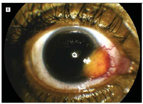
一例人类免疫缺陷病毒阳性患者发生的鸟分枝杆菌性复杂结膜肉芽肿病
一位46岁的HIV阳性男性患者,角膜和结膜之间出现不断扩大的无痛肿块(A)。组织活检显示纤维血管组织和众多分枝杆菌(齐-尼二氏抗酸染色,原始放大倍数×100)(B)。疾病预防和控制中心的物种形成测试显示病原体为鸟分支杆菌复合群。
标题:Mycobacterium avium Complex Conjunctival Granuloma in a Human Immunodeficiency Virus–Positive Patient
出处:Arch Ophthalmol. 2011. 129(12):1609.
作者:Bromley JG, Woodward MA, Grossniklaus HE
A 46-year-old human immunodeficiency virus–positive man had an enlarging, painless limbal conjunctival mass (A), and biopsy of the lesion showed fibrovascular tissue with numerous mycobacteria (Ziehl-Neelsen stain, original magnification ×100) (B). Speciation performed by the Centers for Disease Control and Prevention revealed the pathogen to be mycobacterium avium complex.
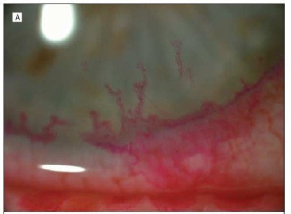
原发单纯疱疹性角膜缘部炎(求指导下,标题翻译为什么比较准确)
一位70岁的女性患者,上下睑缘溃疡穿孔,图示经典的树枝状溃疡边缘。血清中单纯疱疹IgG抗体滴度是正常上限的30倍。
标题:Primary Herpes Simplex Blepharitis and Limbal Keratitis
作者:Allon Barsam, MD, MRCOphth; Eric D. Donnenfeld, MD; Henry D. Perry, MD
出处:Arch Ophthalmol. 2012;130(2):179-179.
A 70-year-old woman with punched-out ulcerations on upper and lower lid margins and classical dendrites (A) encircling the limbus (B). Herpes simplex IgG serum antibody titers were 30 times the upper limit of normal.
YAG激光玻璃体切除治疗Valsalva视网膜病变的玻璃体膜下出血。
一位25岁的患者突发视物不清。A.视力检查时左眼为60cm数指,眼底可见一处大的玻璃体下出血。B.诊断为Valsalva视网膜病变,行YAG激光玻璃体切除术。C.一个月后复查,出血完全消退,视力恢复到20/20。
标题:YAG Hyaloidotomy of Subhyaloid Hemorrhage in Valsalva Retinopathy
作者:Ekta Rishi, MS; Shalini Singh, MS; Roy Rupak, MS; Ramesh Venkatesh, MS
出处:Arch Ophthalmol. 2012;130(2):170-170.
A 25-year-old man had sudden dimness of vision. A, On examination, visual acuity was counting fingers OD at 60 cm and the fundus had a large subhyaloid hemorrhage mound. B, A diagnosis of Valsalva retinopathy was made and YAG hyaloidotomy was done. C, One month after laser treatment there was complete resolution of the hemorrhage with visual acuity recovery to 20/20.
棘阿米巴角膜炎的表现-放射状角膜神经炎
一名使用月抛隐形眼镜30岁女性,经微生物学证实患有棘阿米巴性角膜炎。眼前节谱域OCT显示放射状角膜神经炎的细节。
标题:Radial Keratoneuritis in Acanthamoeba Keratitis
作者:Ines Samet-Tran, MD; Jonathan Letsch, MD; Benoit Guignier, MD; Tristan Bourcier, MD, PhD
出处:Arch Ophthalmol. 2012;130(3):328-328.
A spectral-domain optical coherence tomography examination was performed with the anterior segment module to provide images detailing radial keratoneuritis in a 30-year-old woman with microbiologically proven Acanthamoeba keratitis who used monthly disposable soft contact lenses.
请点击看大图,另欢迎指出错误和进行补充。
怀疑脉络膜参与的全身性粘膜相关淋巴样组织淋巴瘤
图1.外部检查发现两眼增大的泪腺。
图2.眼眶部MRI显示泪腺团块,沿着其轮廓没有发现骨转移的证据(图A箭头所示)。左眼眼后节值得注意(图B)。
图3.眼后节检查发现多个离散的橙黄色视网膜下团块,弥漫在整个眼底,未发生玻璃体炎。
标题:Systemic MALToma With Presumed Choroidal Involvement
作者:Seongmu Lee, MD; Raymond S. Douglas, MD, PhD
出处:Arch Ophthalmol. 2012;130(3):379-379.
Figure 1. External examination revealed enlarged lacrimal glands on both sides.
Figure 2. Magnetic resonance imaging of the orbit revealed lacrimal gland masses that followed the contour of adjacent structures without evidence of bony erosion (A) (arrowheads); involvement of the left posterior segment was noted (B) (arrow).
Figure 3. Posterior segment examination revealed multiple, discrete, yellow-orange subretinal masses situated diffusely throughout the fundus without vitritis.
回复 5楼:
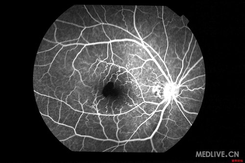
楼主不介意插楼吧![]() ,看到Valsalva视网膜病变想起不久前查资料看到的图,原帖在协和眼科医生论坛。
,看到Valsalva视网膜病变想起不久前查资料看到的图,原帖在协和眼科医生论坛。
患者,男性,20岁,因1天前做俯卧撑后双眼视力突然下降.
视力OD:0.4 OS:0.6 双眼前节(-),眼底,黄斑出血。
另有病例一篇:

地塞米松植入剂迟发迁移至前房
一位女性患者既往存在假晶状体并行玻璃体切割术 ,因视网膜中央静脉阻塞接受眼部地塞米松植入。一个月后,植入物迁移到了前房。角膜水肿持续恶化,亟需通过穿刺术去除易碎的植入物。
Late Migration of Dexamethasone Implant Into Anterior Chamber
Kevin M. Cronin, BA; Kishan Govind, BA; Shree K. Kurup, MD
Arch Ophthalmol. 2012;130(6):711-711.
A woman with pseudophakia (stable, well centered) and a history of vitrectomy received a dexamethasone implant for central retinal vein occlusion. A month later, the implant migrated into the anterior chamber. Worsening preexisting corneal edema mandated removal of the friable implant through a paracentesis.
好吧,这一期的两篇都是植入剂
地塞米松玻璃体植入剂脱位
一名46岁的女性在骑自行车后出现视力障碍。左眼假晶状体(巩膜固定后房型人工晶体)在2周前接受了地塞米松玻璃体植入(Ozurdex)。由于慢性中间葡萄膜炎,右眼已盲。左眼视力 20/100,并且在ETDRS视力表上比植入前还差2行。左眼出现角膜水肿,植入剂移入前房。左眼眼压10mmHg。在移除植入物后,角膜水肿改善,视力慢慢恢复。
Dislocation of Dexamethasone Intravitreous Implant
Bogomil Voykov, MD, FEBO; Karl Ulrich Bartz-Schmidt, MD
Arch Ophthalmol. 2012;130(6):706-706.
A 46-year-old woman had visual acuity impairment in the left eye after riding a bicycle. The pseudophakic left eye (scleral-fixated posterior chamber intraocular lens) had received a dexamethasone intravitreous implant (Ozurdex) 2 weeks earlier. Visual acuity was 20/100 OS and was 2 lines worse on the Early Treatment Diabetic Retinopathy Study chart than before implantation. The right eye was blind due to chronic intermediate uveitis. On examination, distinct corneal edema of the left eye was seen. The implant was dislocated in the anterior chamber with endothelial contact. Intraocular pressure was 10 mm Hg OS. After explantation of the implant, both corneal edema and visual acuity slowly recovered.